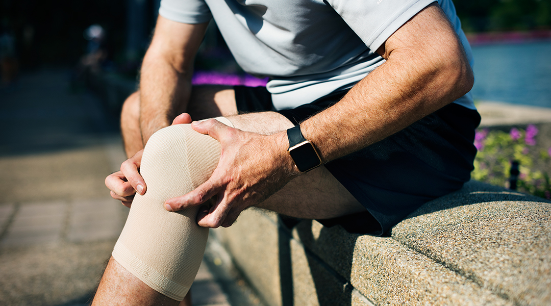


Accidents can happen at any time, and even a simple slip can lead to serious injuries. In this article, we explore a real-life medical case of a patient who experienced pain and swelling in the left forearm after a fall. We’ll walk through what happened, how doctors responded, and how surgery helped restore function and healing.
Case Overview
- Presenting Complaint: Pain and swelling in the left forearm
- Cause: Accidental slip and fall while walking
- Diagnosis: Left forearm fracture
- Treatment: Surgical fixation with plates under regional anesthesia
What Happened to the Patient?
The patient reported sudden pain and swelling in the left forearm after slipping and falling while walking. There was no history of head injury, bleeding from the ears, nose, or throat (ENT bleed), or loss of consciousness(LOC), which are commonly checked to rule out serious trauma.
The main concern was localized to the left forearm, where the patient had difficulty moving it due to pain and swelling.
Initial Medical Examination
Once the patient was brought to the hospital, doctors carried out a general and systemic examination.
Vital Signs and Observations:
- Consciousness: Patient was alert and responded to verbal commands
- Temperature: 98°F (normal)
- Pulse Rate (PR): 96 beats per minute
- Blood Pressure (BP): 110/70 mmHg (within normal range)
- Oxygen Saturation (SpO2): 97% on room air
General Condition
- No signs of jaundice (icterus)
- No cyanosis (bluish color of skin due to lack of oxygen)
- No clubbing (enlarged fingertips often seen in chronic illnesses)
- No lymph node swelling (lymphadenopathy)
Systemic Examination
- Cardiovascular System (CVS): Heart sounds S1 and S2 were normal
- Respiratory System (RS): Breath sounds were clear on both sides
- Abdomen (P/A): Soft and non-tender
- Central Nervous System (CNS): No findings suggestive of neurological damage
Forearm-Specific Findings
The injury was mainly located in the left forearm, with the following signs:
- Tenderness: Present
- Swelling: Present
- Range of Motion (ROM): Painful and restricted
- DNVD (Distal Neurovascular Deficit): Not present (nerves and blood flow to the hand were intact)
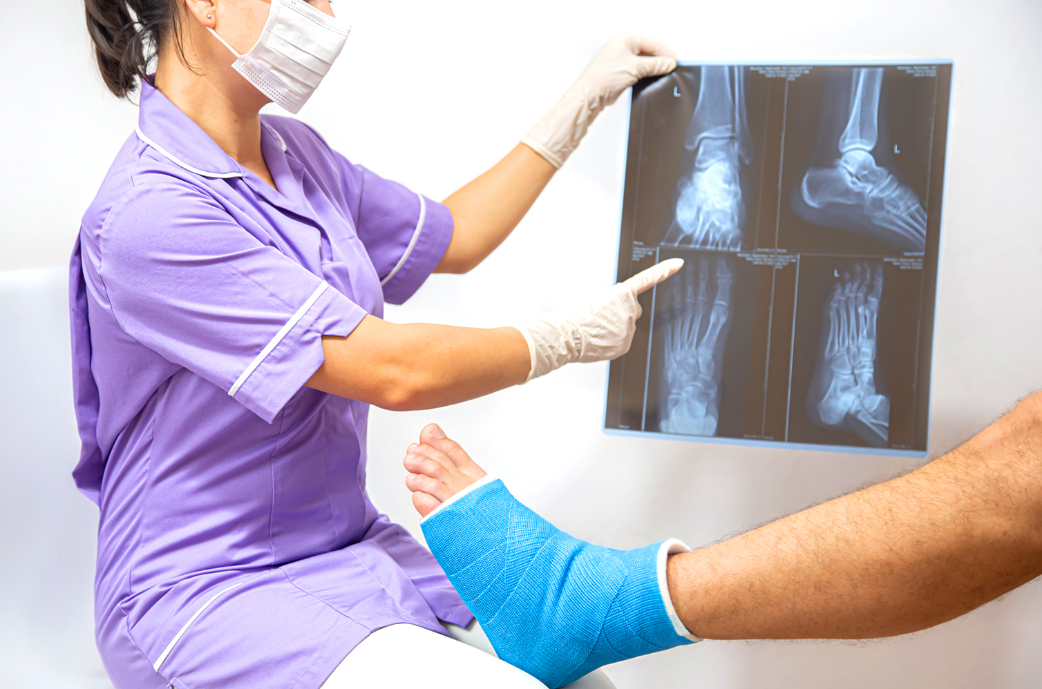
Diagnosis: Fracture of the Left Forearm
Based on clinical findings and further imaging (such as an X-ray), a fracture was confirmed in the bones of the left forearm: radius and ulna.
These are the two long bones in the forearm. A break in either or both can affect the arm’s function and needs careful treatment.
Treatment Plan: Surgery for Bone Fixation
Given the severity of the fracture and reduced movement, doctors decided that surgical intervention was the best course of action.
The goal was to:
- Realign the bones properly (called fracture reduction)
- Fix them in place using metal plates
- Ensure healing in the correct position
- Prevent complications such as bone deformity or loss of function
Surgery Procedure Explained
Type of Anesthesia:
- Regional anesthesia (block): Only the arm was numbed; the patient was awake but did not feel pain in the affected area.
Positioning and Preparation:
- The patient was placed in a supine position (lying on the back).
- The left forearm was placed on an arm board for support.
- The area was cleaned, sterilized, and draped.
Surgical Steps:
1. Volar Approach to Radius (Front Side of Forearm)
- A 5 cm incision was made over the front (volar) side of the forearm.
- The skin and underlying tissue were gently separated.
- The fracture site of the radius bone was exposed.
- The bone was realigned (reduced) and fixed using a 5-hole metal plate.
2. Ulnar Approach (Back/Dorsal Side of Forearm)
- A second incision was made over the ulnar side of the forearm.
- The fracture on the ulna bone was also exposed.
- It was reduced and fixed using an 8-hole plate.
Finishing Steps:
- Surgeons ensured the bones were correctly aligned using C-arm X-ray guidance.
- A thorough wound wash was given to reduce infection risk.
- The skin was closed with sutures.
- A dressing was applied to keep the area sterile and support healing.
Post-Surgery Recovery and Care
After surgery, the patient was moved to the recovery ward and monitored for:
- Pain control
- Bleeding or infection
- Swelling
- Signs of proper healing
A cast or splint may be used to protect the area, and the patient will likely begin physical therapy once healing begins to restore movement and strength.
Importance of Timely Treatment
Forearm fractures might seem like a minor injury, but if not treated properly, they can lead to:
- Permanent deformity
- Loss of arm function
- Nerve or vessel damage
- Chronic pain
Timely surgical treatment, as done in this case, helps:
- Ensure faster and correct healing
- Restore full function
- Prevent complications
What Is Plate Fixation in Fractures?
Plate fixation is a surgical method where metal plates and screws are used to hold broken bones in the correct position. It is often used for fractures that:
- Involve multiple parts
- Are misaligned
- Do not respond well to plaster or casting alone
The plates stay in place permanently or may be removed later based on the patient’s progress.
How to Prevent Falls and Forearm Injuries
- Wear proper footwear with a grip
- Keep walkways dry and clutter-free
- Use handrails when walking down stairs
- Be mindful of slippery or uneven surfaces
- Avoid walking while distracted or on your mobile phone
Conclusion
This case highlights the importance of seeking immediate medical help after a fall. Even if the injury appears minor, hidden fractures can worsen without proper care. Thanks to a detailed surgical approach, the patient’s forearm was successfully treated using metal plates, allowing a clear path toward recovery.
If you or someone you know has experienced a similar injury, do not delay medical evaluation. Prompt care can make all the difference in healing and quality of life.
FAQs
1. Is surgery always needed for forearm fractures?
No. Minor fractures may heal with a cast. Surgery is required when bones are displaced or broken in multiple places.
2. How long does it take to recover after forearm fracture surgery?
Generally, 6 to 8 weeks for the bone to heal, but full strength and motion may take up to 3 to 6 months with proper physiotherapy.
3. Will the metal plates be removed?
Sometimes they are left permanently. In younger patients or if they cause discomfort, they may be removed after healing.